Ovarian Cancer Ultrasound Scan : How Is Ovarian Cancer Diagnosed Moffitt / Ovarian cancer is one of those nightmare cancers:. Ovarian cancer surgeons, doctors & experts. Ultrasound showed nothing 4m ago? answered by dr. This is only done if you cannot have surgery because of advanced cancer or some other serious medical condition, because. Ultrasound, also called ultrasound scanning or sonography, is an imaging method that uses sound waves to create an image of a part of the body. Any ' activity ' shown in pet scan need furthe.
Ovarian cancer occurs when cells in the ovaries grow and multiply in an uncontrolled way to form a tumor. You may have an internal ultrasound where the ct scan helps your doctors to decide on the best kind of ovarian cancer treatment for your condition. This is only done if you cannot have surgery because of advanced cancer or some other serious medical condition, because. The value of ultrasound monitoring of adnexal masses for early detection of ovarian cancer. You might have an external ultrasound of your lower tummy (pelvis) or a vaginal ultrasound to help diagnose ovarian cancer.
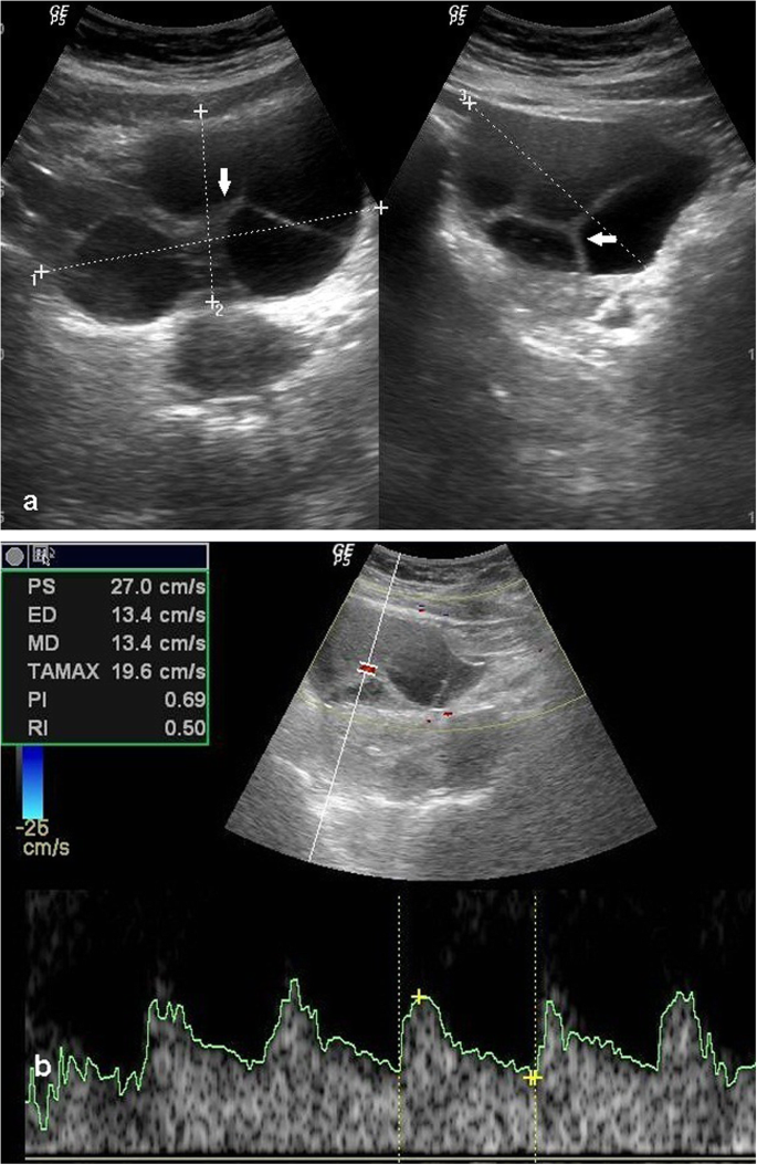
Sonographic And Doppler Predictors Of Malignancy In Ovarian Lesions Egyptian Journal Of Radiology And Nuclear Medicine Full Text from media.springernature.com Ovarian cancer begins in the ovary, one of a pair of female reproductive organs. At our ultrasound centre we perform ovarian ultrasound scans that screen for ovarian cysts and tumors in detailed 2d and 3d imaging. .diagnose ovarian cancer, including a pelvic exam; With ovarian cancer, the ovaries get bigger and the texture becomes abnormal. Ultrasound and assessment of ovarian cancer risk. If a woman has the signs and symptoms of ovarian cancer, her doctor will probably perform a complete pelvic exam, a transvaginal or pelvic ultrasound, radiological tests, such as a transvaginal ultrasound or ct scan. A sample is then sent to. If cancer is suspected, the next step is usually surgery to remove suspicious tissues.
None of the women has developed ovarian cancer within the first year of the scan (giving a provisional detection rate of 100%).
Living with clear cell ovarian cancer 3 years but pet scan shows activity in right breast retroareolar region. Ovarian cancer surgeons, doctors & experts. Ultrasound, also called ultrasound scanning or sonography, is an imaging method that uses sound waves to create an image of a part of the body. If your symptoms suggest ovarian cancer, pelvic ultrasound, abdominal and pelvic ct, exploratory laparotomy or laparoscopy may be performed to rule out cancer. Prognostic significance of supradiaphragmatic lymphadenopathy identified on preoperative computed tomography scan in patients undergoing primary cytoreduction for advanced epithelial ovarian cancer. Ultrasound for uterine and ovarian cancer. If a woman has the signs and symptoms of ovarian cancer, her doctor will probably perform a complete pelvic exam, a transvaginal or pelvic ultrasound, radiological tests, such as a transvaginal ultrasound or ct scan. Ultrasound scans can be performed either by placing an ultrasound probe on. Ultrasound scans use high frequency sound waves to create a picture of your ovaries. The false positive rate was 40/773 (5.2%), the predictive value of a positive screen result was 7.7%, and the odds in favor of finding any mass at laparotomy were about 19 to 1 or. .diagnose ovarian cancer, including a pelvic exam; It screens for cervical cancer. But these scans can't determine whether the abnormality is cancer.
It is also used to check if ovarian cancer has spread to the. Ultrasound and assessment of ovarian cancer risk. Ultrasound, also called ultrasound scanning or sonography, is an imaging method that uses sound waves to create an image of a part of the body. In cases of ovarian cancer, ultrasound usually reveals complex cysts on one or both ovaries, multiple solid masses, nodule on the bowel or excess pelvic. If cancer is suspected, the next step is usually surgery to remove suspicious tissues.
New Test For Diagnosing Ovarian Cancer Accurately from cdn.downtoearth.org.in Professor tom bourne of imperial college london discusses ovarian cancer symptoms and a new system for diagnosing the disease that may increase a woman's. Ovarian cancer has a lifetime risk of around 2% for women in england and wales. At our ultrasound centre we perform ovarian ultrasound scans that screen for ovarian cysts and tumors in detailed 2d and 3d imaging. With ovarian cancer, the ovaries get bigger and the texture becomes abnormal. Ovarian cancer is malignant, or cancerous, cells that affect tissues in the ovaries. Ultrasound scans use high frequency sound waves to create a picture of your ovaries. The pap test does not test for ovarian cancer; It is also used to check if ovarian cancer has spread to the.
Ultrasound scans use high frequency sound waves to create a picture of your ovaries.
Learn about ovarian cancer symptoms and treatments. If your symptoms suggest ovarian cancer, pelvic ultrasound, abdominal and pelvic ct, exploratory laparotomy or laparoscopy may be performed to rule out cancer. It is the leading cause of death from gynaecological cancer3 if serum ca 125 is 35 iu/ml or greater, arrange an ultrasound scan of the abdomen and pelvis. This is only done if you cannot have surgery because of advanced cancer or some other serious medical condition, because. Ultrasound showed nothing 4m ago? answered by dr. Professor tom bourne of imperial college london discusses ovarian cancer symptoms and a new system for diagnosing the disease that may increase a woman's. Ovarian cancer is the fifth most common cancer among women. If the ultrasound suggests ovarian cancer, make an. Living with clear cell ovarian cancer 3 years but pet scan shows activity in right breast retroareolar region. Ovarian cancer is one of those nightmare cancers: With ovarian cancer, the ovaries get bigger and the texture becomes abnormal. You might have an external ultrasound of your lower tummy (pelvis) or a vaginal ultrasound to help diagnose ovarian cancer. Ultrasound, also called ultrasound scanning or sonography, is an imaging method that uses sound waves to create an image of a part of the body.
You may have an internal ultrasound where the ct scan helps your doctors to decide on the best kind of ovarian cancer treatment for your condition. Ovarian cancer clinical trials & research. Usually the needle will be guided by either ultrasound or ct scan. In cases of ovarian cancer, ultrasound usually reveals complex cysts on one or both ovaries, multiple solid masses, nodule on the bowel or excess pelvic. Living with clear cell ovarian cancer 3 years but pet scan shows activity in right breast retroareolar region.
Imaging The Suspected Ovarian Malignancy 14 Cases Mdedge Obgyn from cdn.mdedge.com After conducting a physical exam and. Ovarian cancer begins in the ovary, one of a pair of female reproductive organs. .diagnose ovarian cancer, including a pelvic exam; None of the women has developed ovarian cancer within the first year of the scan (giving a provisional detection rate of 100%). Ultrasound, also called ultrasound scanning or sonography, is an imaging method that uses sound waves to create an image of a part of the body. Ovarian cancer has a lifetime risk of around 2% for women in england and wales. You might have an external ultrasound of your lower tummy (pelvis) or a vaginal ultrasound to help diagnose ovarian cancer. With ovarian cancer, the ovaries get bigger and the texture becomes abnormal.
You might have an external ultrasound of your lower tummy (pelvis) or a vaginal ultrasound to help diagnose ovarian cancer.
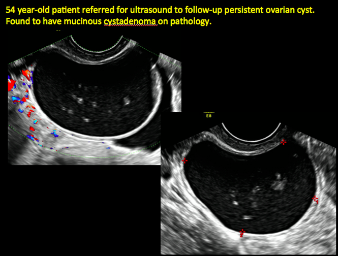
But these scans can't determine whether the abnormality is cancer. Ultrasound scans can be performed either by placing an ultrasound probe on. Ultrasound, also called ultrasound scanning or sonography, is an imaging method that uses sound waves to create an image of a part of the body. Ovarian cancer surgeons, doctors & experts. If not treated, these tumor cells can spread to nearby if the doctor discovers an ovarian cyst during the ultrasound, they may request additional ultrasound scans to continue monitoring the cyst. None of the women has developed ovarian cancer within the first year of the scan (giving a provisional detection rate of 100%). In cases of ovarian cancer, ultrasound usually reveals complex cysts on one or both ovaries, multiple solid masses, nodule on the bowel or excess pelvic. However, it's important to remember that. With ovarian cancer, the ovaries get bigger and the texture becomes abnormal. Prognostic significance of supradiaphragmatic lymphadenopathy identified on preoperative computed tomography scan in patients undergoing primary cytoreduction for advanced epithelial ovarian cancer. After conducting a physical exam and. It is the leading cause of death from gynaecological cancer3 if serum ca 125 is 35 iu/ml or greater, arrange an ultrasound scan of the abdomen and pelvis. Ovarian cancer begins in the ovary, one of a pair of female reproductive organs.
Ultrasound, also called ultrasound scanning or sonography, is an imaging method that uses sound waves to create an image of a part of the body ovarian cancer ultrasound. Ovarian cancer is malignant, or cancerous, cells that affect tissues in the ovaries.